Introduction
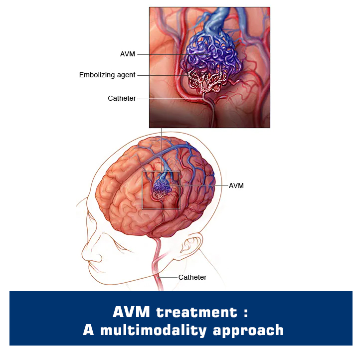
AVM or arteriovenous malformation is an entity of the brain where arteries and vein form irregular connections between them called nidus instead of a normal capillaries network. Presentation of AVM varies from seizures, headache and bleeding to asymptomatic. Treatment or better management of AVM is based upon the presentation and angio-architecture.
Primary goal of AVM treatment
The primary goal of treatment depends upon the type of presentation. For example, if patient presents with a bleed, then the goal is to prevent rebleed. Likewise, if a patient presents with seizures and on DSA no weak point or other weak angio-architecture is noted then it’s better to manage on medications.
Indications for AVM surgery or other forms of treatment
AVM is categorized into different grades. One of the most commonly used classifications is Spetzer Martin Grading, although there are other classifications also. Depending on the grading, location and presentation patient will be offered only surgical excision, AVM embolization or gamma knife or a combination of two or all. That’s why AVM treatment is a multimodality treatment.
Patients prepare for AVM treatment
Preoperative preparation for AVM surgical excision or AVM Embolization (Endovascular) involves a thorough evaluation, which includes medical history review, physical examination, and imaging studies (NCCT head, MRI Head and Cerebral DSA). Based on all the available investigations and history a treatment plan will be charted. Sometimes in addition to usual surgical excision or endovascular embolization, a patient may need EVD/VP shunt for hydrocephalus.
Procedure performed for AVM treatment
AVM excision or embolization is typically performed under general anaesthesia. An incision is made along the scalp. A surgical microscope is commonly used during the procedure to provide a magnified view, improving precision and minimizing the risk of damage to surrounding structures.
In embolization, only a small needle puncture is done commonly in the groin (femoral artery) and sometimes in the wrist (radial artery) and the procedure is performed with small calibre catheters and wires in Neuroangio suite.
Gamma knife is a form of radiation and can be performed in a conscious state without general anaesthesia.
Difference between AVM excision and AVM embolization
AVM excision and AVM embolization are two different approaches to treat AVM. Surgical excision involves the surgical removal of AVM nidus along with occlusion of feeding arteries and draining veins, while AVM embolization involves the occlusion of AVM nidus along with feeding arteries and veins and is sometimes followed by surgical excision also. The choice between the two procedures depends on various factors, including the patient's overall health, anatomy, and surgeon's expertise.
The third option of Gamma knife will also be considered as a standalone treatment or in combination of the above treatments.
Postoperative care involves and the long-term outcomes
Post-surgery or AVM embolization patient is shifted to NSICU for neurological and hemodynamic monitoring. Patients may have break through bleeding or seizures in a post-op period for which medical optimization will be done. Long-term and regular follow-up is needed post-AVM treatment.
Conclusion:
AVM management is a highly complex, even more individualistic and algorithmic multimodality treatment where experts from various fields discuss outcomes of a single or combination of treatments to reach at a logical conclusion which will be best for a patient in the long term.
Shushrut Brain and Spine has developed an AVM Center in order to offer patients individualized treatment. As a leader in diagnosing, researching and treating vascular anomalies and vascular tumors, our team of specialists provide comprehensive treatment and care.