



Brain Aneurysm: Symptoms, Causes, Diagnosis and Treatment

Quick answer

A brain aneurysm is a weakened bulge on the wall of a brain artery. Most unruptured aneurysms produce no symptoms and are found on imaging done for another reason. A ruptured aneurysm typically presents with a sudden, severe, unlike any previous headache, often with vomiting, neck stiffness, brief loss of consciousness or focal neurological signs, and is a medical emergency.

Show More

What is a brain aneurysm?

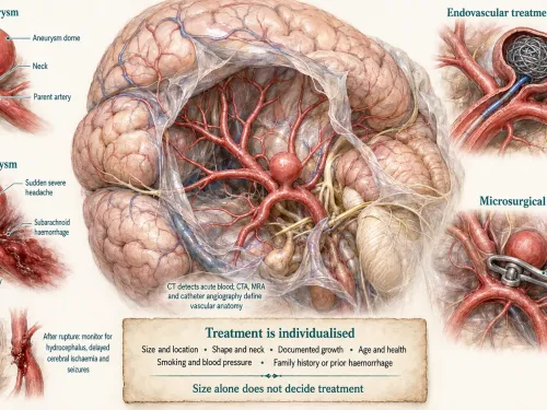
An intracranial or cerebral aneurysm forms when part of an artery wall becomes weakened and bulges. The common saccular aneurysm looks like a small pouch arising from a vessel. Some aneurysms are fusiform, involving a longer segment of the artery. The clinical risk is not simply the presence of the bulge; doctors assess the possibility of growth or rupture, the consequences of treatment and the patient’s individual risk profile.

Symptoms: unruptured and ruptured aneurysms are very different

Unruptured aneurysm

Most small unruptured aneurysms are silent and are found incidentally on CT angiography or MR angiography. When an aneurysm becomes large or presses on nearby nerves, symptoms can include pain around or behind an eye, double vision, visual change, a dilated pupil, facial numbness or other focal neurological symptoms. These are not specific and require imaging to identify the cause.

Ruptured aneurysm

When an aneurysm ruptures, blood often enters the subarachnoid space around the brain. The immediate problems are the haemorrhage itself and the risk of rebleeding. Over the following days, patients can also develop hydrocephalus, delayed cerebral ischaemia, electrolyte disturbances, seizures and other complications. Treatment therefore involves both securing the aneurysm and managing the consequences of the bleed in a neurocritical-care setting.

Why do aneurysms form?

Aneurysm formation is usually multifactorial. Age, high blood pressure and cigarette smoking are important modifiable factors. Family history can matter, particularly when more than one close relative has had an intracranial aneurysm or aneurysmal subarachnoid haemorrhage. Certain inherited connective-tissue or vascular disorders are associated with higher risk. Many patients, however, have no single identifiable cause.

How is an aneurysm diagnosed?

In suspected subarachnoid haemorrhage, non-contrast CT of the head is often the first emergency test because it can rapidly detect acute blood. CT angiography can then demonstrate the aneurysm and vascular anatomy. Depending on timing and findings, additional tests may be required. Catheter cerebral angiography provides high-resolution information about vessel anatomy and is often used for treatment planning. MR angiography is useful in selected non-emergency situations and for follow-up.

Does every unruptured aneurysm need treatment?

No. For an unruptured aneurysm, the decision balances the estimated lifetime rupture risk against the immediate and long-term risks of intervention. Size is relevant but not the only variable. Location, shape, irregularity, documented growth, age, smoking, blood pressure, family history, prior subarachnoid haemorrhage and the technical features of treatment all influence the recommendation. Some aneurysms are monitored with interval imaging and risk-factor control.

Treatment options

Endovascular treatment

Endovascular procedures reach the aneurysm from within the blood vessels, commonly through an artery in the wrist or groin. Coils can be placed inside the aneurysm to promote thrombosis. Wide-neck or complex aneurysms may require balloon-assisted or stent-assisted techniques. Flow-diverting stents are used for selected aneurysms whose anatomy is less suited to conventional coiling. Device choice depends on anatomy and whether the aneurysm has ruptured, because antiplatelet medication may be required with some stents.

Microsurgical clipping

Clipping is performed through a craniotomy. The neurosurgeon exposes the aneurysm and places a clip across its neck to exclude it from circulation while preserving the parent artery and important branches. Clipping may be particularly suitable for certain aneurysm locations or configurations, when a durable direct reconstruction is desirable, or when associated clot or brain compression also needs surgical treatment.

Observation and risk-factor management

When observation is selected, blood pressure control and smoking cessation are important. Follow-up imaging may be recommended to detect growth or morphological change. The interval is individualized; a rigid schedule is not appropriate for every aneurysm.

Why both surgical and endovascular perspectives matter

Modern aneurysm care is not a competition between clipping and coiling. Some aneurysms are clearly better suited to one route; others are genuinely debatable. Reviewing the same angiographic anatomy from microsurgical and endovascular perspectives helps patients understand the trade-offs, including procedural risk, durability, need for antiplatelet medication and follow-up imaging.

Recovery after rupture

Securing the aneurysm is only one part of recovery from subarachnoid haemorrhage. Patients may need intensive neurological monitoring for vasospasm and delayed ischaemia, management of hydrocephalus, respiratory or cardiac complications, seizure assessment and later rehabilitation. Fatigue, cognitive slowing and emotional change can persist even when limb strength appears normal.

What happens after a ruptured aneurysm is secured?

The danger does not end once the aneurysm is clipped or coiled. During the following days, the brain remains vulnerable to delayed cerebral ischaemia, hydrocephalus, seizures and systemic complications. Patients may need repeated neurological examinations, vascular monitoring, treatment to maintain appropriate circulation and drainage of cerebrospinal fluid if pressure builds. This is why ruptured aneurysm care belongs in a setting with neurocritical-care capability as well as procedural expertise.

How doctors estimate risk in an unruptured aneurysm

No single measurement determines rupture risk. Doctors consider maximum diameter, site in the cerebral circulation, irregular shape or daughter sacs, interval growth, previous subarachnoid haemorrhage and patient factors such as age, smoking and hypertension. Family history and certain inherited disorders can also influence the discussion. At the same time, treatment risk rises with some complex anatomies and medical conditions. The decision is therefore a comparison of two imperfect estimates: the risk of leaving the aneurysm untreated and the risk of treating it now.

Lifestyle changes matter even when no procedure is planned

Smoking cessation and blood-pressure control are among the most practical modifiable measures. Patients should also take prescribed cardiovascular medication consistently and avoid assuming that supplements can reduce aneurysm size. Normal physical activity is often possible with an unruptured aneurysm, but individualized advice is appropriate when the aneurysm is large, symptomatic or awaiting treatment.

Screening is selective, not universal

People sometimes request an angiogram because they have headache. Routine aneurysm screening for the general population is not recommended simply on that basis. Screening discussions are more relevant when there is a strong family pattern, particularly multiple close relatives with aneurysm or subarachnoid haemorrhage, or an associated inherited condition. Before screening, it is worth discussing what an incidental small aneurysm would mean because discovery can lead to years of surveillance and difficult risk decisions.

Recovery after subarachnoid haemorrhage can be deceptively slow

A patient may regain full limb strength yet continue to experience fatigue, slowed processing, poor concentration, sleep disturbance, anxiety or reduced exercise tolerance. Families can mistake this for lack of effort because the deficits are less visible than paralysis. Neuropsychological and rehabilitation support may be useful. Return to work and driving should be individualized according to neurological recovery, seizures and local regulations.

What information to carry for a second opinion

For an unruptured aneurysm, bring the CTA or MRA images, catheter angiography if performed, prior imaging for growth comparison, current blood-pressure treatment and details of family history. For a previously treated aneurysm, include the procedural report and follow-up angiograms. Those records often answer more than a new standalone scan.

Blood pressure, smoking and aneurysm growth

Blood pressure control is not merely general health advice in a patient with an aneurysm. Persistent hypertension increases haemodynamic stress on arterial walls, while smoking is associated with aneurysm formation and rupture. Stopping tobacco and treating hypertension do not make an existing aneurysm disappear, but they reduce modifiable vascular risk and are part of responsible observation as well as post-treatment care.

Pregnancy and other special situations

Pregnancy, planned major surgery for another condition, long-term anticoagulation or antiplatelet needs can complicate management of an unruptured aneurysm. These situations do not create a universal rule for treatment, but they may change timing and the balance between clipping, coiling, device-based treatment and surveillance. The neurovascular team should coordinate with the relevant obstetric, cardiac or surgical specialists rather than making the aneurysm decision in isolation.

Why follow-up imaging should use comparable studies

Aneurysm growth can be subtle. Comparing studies performed with similar technique and reconstructing the same views is more useful than relying on a one-millimetre difference quoted from separate reports. Apparent change can reflect measurement variation. When true growth is confirmed, however, it can materially alter the treatment discussion because it suggests biological instability.

Headache after a treated aneurysm should still be assessed by pattern

A patient who has undergone coiling or clipping may later develop ordinary migraine or tension-type headache. Not every headache means recurrence. A new thunderclap pattern, new neurological deficit or significant change from baseline deserves urgent reassessment, while stable recurrent headache may be evaluated more routinely. The prior aneurysm history lowers the threshold for taking a major change seriously but should not make every headache an emergency.

Common Patient Questions

Can an aneurysm exist without symptoms?

Yes. Many unruptured aneurysms are incidental findings.

Can a small aneurysm rupture?

Yes, although rupture risk varies. Size is one factor among several, so treatment should not be based on a size threshold alone.

Is a thunderclap headache always an aneurysm?

No, but it is a medical emergency because subarachnoid haemorrhage and other dangerous vascular causes must be excluded quickly.

Can aneurysms run in families?

They can. A strong family history may change screening discussions, especially when multiple close relatives are affected.

Will coiling always avoid open surgery?

Coiling is minimally invasive, but not every aneurysm is suitable. Anatomy and clinical condition determine the approach.

Does clipping mean no future follow-up?

Clipping can be very durable, but follow-up needs depend on the aneurysm, completeness of occlusion and whether other aneurysms are present.

For patients in Noida and the wider region

For suspected aneurysm rupture in Noida, Greater Noida, Ghaziabad or Delhi NCR, the priority is the nearest facility capable of emergency brain imaging, neurovascular treatment and neurocritical care rather than travelling long distances. For stable unruptured aneurysms, patients from UP, MP or Bihar can bring CTA/MRA images and any catheter angiography for planned multidisciplinary review.

Editorial medical references

  • NINDS: Cerebral Aneurysms
  • AANS: Cerebral Aneurysm

A common misconception

A brain aneurysm is not the same as a brain haemorrhage. It is the underlying weak point on an artery. A haemorrhage occurs only when the aneurysm ruptures or leaks.

Do not find what you're looking for?

Our specialists are here to help. Get expert answers tailored to your condition—no waiting, no guesswork