

Pituitary Tumour Treatment: How the Appropriate Option Is Chosen

Quick answer

Pituitary tumour treatment is chosen after considering tumour size, hormonal activity, effect on vision and adjacent structures, and the patient's overall condition. Some tumours are safely observed with periodic imaging and hormonal review. Some prolactinomas respond well to medication as first-line treatment. Others, particularly those causing visual field loss or hormonal excess resistant to medication, are treated surgically, most commonly by an endoscopic endonasal approach.

Show More

Image
Pituitary Tumour Treatment


Pituitary tumours are not all treated the same way

Most pituitary tumours are adenomas and are not cancers. Their importance comes from two potential effects: pressure on nearby structures, particularly the optic chiasm, and abnormal hormone production or loss of normal pituitary function. A small hormone-secreting tumour can be clinically important even when it is only a few millimetres, while a larger nonfunctioning lesion may present mainly through visual loss.

The first step is a combined endocrine, visual and imaging assessment

  • MRI of the pituitary region, usually with contrast and dedicated sellar sequences.
  • Pituitary hormone testing to identify hypersecretion and hormone deficiency.
  • Formal visual-field testing when the lesion approaches or compresses the optic nerves or chiasm.
  • Assessment of headache, eye movements and other neurological symptoms.
  • Review of medications and physiological states that can alter hormone levels.

Observation for selected incidental tumours

A small incidentally found lesion that does not produce excess hormones, does not threaten vision and has no concerning growth pattern may be monitored. Surveillance typically includes clinical review and interval MRI, with endocrine or visual testing depending on size and proximity to the optic pathways. The schedule is individualized and can become less frequent when stability is established.

Medication can be the primary treatment for some functioning tumours

Prolactinoma

Prolactin-secreting adenomas often respond to dopamine-agonist medication, which can lower prolactin and shrink the tumour. Surgery may still be considered in selected patients with medication intolerance or resistance, specific anatomical features, rapidly progressive visual compromise in certain circumstances, or after a shared discussion of long-term options. The important point is that a prolactinoma should not automatically be sent for surgery before its endocrine diagnosis is established.

Other functioning adenomas

Growth-hormone and ACTH-secreting adenomas commonly have surgery as a central treatment when resectable, with medication and radiation used according to postoperative hormone status, residual tumour and disease control. TSH-secreting tumours also require endocrine-directed planning. The biochemical goal is as important as the anatomical MRI result.

When surgery is considered

  • Visual-field loss or other neurological compromise from tumour compression.
  • A lesion abutting or compressing the optic apparatus where growth poses a visual risk.
  • Pituitary apoplexy with significant visual or neurological deterioration.
  • Functioning tumours for which surgery is the appropriate disease-specific treatment.
  • Progressive enlargement of a nonfunctioning tumour with mass effect.
  • Selected prolactinomas after individualized endocrine and surgical discussion.

How pituitary surgery is performed

Most surgically treated pituitary adenomas are approached through the nose and sphenoid sinus using an endoscopic or microscopic transsphenoidal route. This reaches the gland from below without a conventional craniotomy. The aim is to remove the tumour while preserving normal pituitary tissue, the optic apparatus and the major arteries around the sella. Very large, irregular or laterally extending lesions occasionally require a different or combined strategy.

What makes pituitary surgery different from other brain tumour surgery

Success is judged by more than how much tissue is removed. Hormone normalization, visual recovery, preservation of normal pituitary function and avoidance of cerebrospinal-fluid leakage or vascular injury are key outcomes. A postoperative MRI may show an excellent anatomical resection while endocrine disease persists, or hormone control can be achieved even when tiny radiological residual tissue remains.

Role of radiation and radiosurgery

Focused radiosurgery or fractionated radiotherapy can be used for selected residual or recurrent tumours, especially when further surgery is unlikely to provide adequate control or when hormonal disease remains active. The effect can take time, and long-term pituitary hormone deficiency is an important consideration. Radiation therefore forms part of a longitudinal plan rather than an automatic next step for every residual tumour.

Pituitary apoplexy is an emergency pattern

Why multidisciplinary care is particularly important

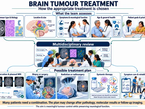
Pituitary disease sits at the boundary of endocrinology, neurosurgery, ophthalmology and radiology. A coordinated plan avoids two common mistakes: treating a hormone disorder as if it were only an MRI finding, and focusing on hormone levels while missing visual or neurological compression. Long-term follow-up may continue even after successful surgery because pituitary function can change over time.

Hormone symptoms can be the first clue

Excess prolactin can cause menstrual disturbance, infertility, reduced libido or galactorrhoea. Growth-hormone excess can produce enlargement of hands and feet, facial change, sweating, sleep apnoea, diabetes and hypertension. ACTH excess can cause features of Cushing disease. These symptoms can develop gradually and may lead to diagnosis before visual problems occur. A pituitary lesion therefore needs biochemical classification, not just size measurement.

Hormone deficiency also matters

A large tumour can compress normal pituitary tissue and reduce cortisol, thyroid or gonadal hormone production. Cortisol deficiency is particularly important because it can become medically dangerous during illness or surgery. Preoperative endocrine testing identifies deficiencies that need replacement and prevents the misconception that normal-looking blood tests from a general panel are sufficient.

Visual-field testing can reveal damage the patient has not noticed

Compression of the optic chiasm classically affects the outer visual fields, and patients may adapt slowly enough that they do not recognise the loss. Formal perimetry gives an objective baseline and helps determine urgency. After decompression, repeated visual fields can document recovery or identify persistent compression.

What follow-up looks like after surgery

Early care includes monitoring sodium balance, urine output, cortisol status, vision and symptoms of cerebrospinal-fluid leak. Some patients develop temporary diabetes insipidus, while others may need long-term hormone replacement. Postoperative MRI and hormone testing are timed according to tumour type. A single “normal” scan does not replace endocrine follow-up for a functioning adenoma.

Residual tumour can be managed in several ways

If tissue remains in the cavernous sinus or another high-risk area, observation may be appropriate when it is stable and hormonally inactive. Active residual disease can be treated with medication, focused radiation or, less commonly, further surgery. The threshold for intervention depends on growth, hormone control and proximity to the optic pathways.

Pituitary apoplexy requires attention to both neurology and hormones

Bleeding or infarction within a pituitary tumour can cause sudden headache, visual loss and eye-movement palsies. It can also produce acute cortisol deficiency. Treatment therefore includes urgent endocrine stabilization as well as deciding whether surgical decompression is required. The severity of visual and neurological compromise guides the surgical decision.

The distinction between microadenoma and macroadenoma is useful but incomplete

Size categories help describe pituitary tumours, but management depends on more than the one-centimetre boundary. A small ACTH-secreting adenoma can cause severe systemic disease, while a larger nonfunctioning adenoma may remain stable without immediate symptoms. Hormone biology and relationship to the optic chiasm can be more important than size alone.

Pregnancy and fertility require specific planning

Pituitary tumours can affect fertility through prolactin excess or loss of normal gonadal hormones. Pregnancy can also alter the size and behaviour of some pituitary lesions. Women planning pregnancy may need a coordinated endocrine and neurosurgical plan that addresses medication, visual monitoring and the risk of tumour enlargement. Management should be individualized rather than extrapolated from a non-pregnant adult.

Why pathology and endocrine remission are different outcomes

A tumour can be removed completely on MRI yet continue to produce excess hormone if microscopic active tissue remains. Conversely, biochemical remission can occur even when imaging shows a tiny stable residual. Follow-up therefore combines hormone testing, imaging and symptoms. Declaring success from the scan alone can miss persistent endocrine disease.

Long-term hormone replacement may be part of a good outcome

If the tumour or its treatment permanently reduces normal pituitary function, replacement of cortisol, thyroid hormone, sex hormones or desmopressin may be required. Needing replacement does not necessarily mean treatment failed. The aim is safe tumour and hormone control while restoring normal physiology as far as possible through coordinated endocrine care.

Cavernous-sinus extension changes surgical expectations

Some pituitary adenomas extend laterally around the carotid artery into the cavernous sinus. Aggressive removal in this region can risk cranial nerves and the artery. In such cases a planned residual may be safer, with hormone-directed medication or radiation used if needed. Patients should understand when “complete removal” would create more risk than benefit.

Headache may improve, persist or be unrelated

Pituitary tumours can cause headache, but headache is common in the general population and is not always caused by the lesion. Decompression may improve pressure-related headache in some patients, yet migraine can persist after technically successful surgery. Visual and endocrine indications are usually more objective reasons for treatment than headache alone.

Common patient questions

Does every pituitary tumour need surgery?

No. Observation or medication can be appropriate depending on tumour type, size, hormone activity and visual risk.

Why is prolactin checked before surgery?

A prolactinoma often responds to medication, so identifying it can completely change the first-line treatment plan.

Can pituitary surgery affect hormones?

Yes. The goal is to preserve normal gland function, but temporary or permanent hormone deficiencies can occur and require endocrine follow-up.

Will vision recover after decompression?

Many patients improve, particularly when compression is treated before prolonged severe optic damage, but recovery is not guaranteed.

Is transsphenoidal surgery “brain surgery”?

It is skull-base neurosurgery performed through the nasal corridor to reach the pituitary. It avoids opening the skull in the conventional way for most adenomas.

Can a pituitary tumour return?

Residual or recurrent tumour can occur depending on type and invasiveness, which is why imaging and endocrine follow-up are important.

For patients in Noida and the wider region

Patients in Noida and Delhi NCR with a pituitary lesion should ideally have MRI, pituitary hormone results and formal visual fields reviewed together. Patients travelling from UP, MP or Bihar should bring all endocrine reports, old MRIs and details of any hormone medication so that the team can distinguish observation, medical therapy, surgery and radiation without unnecessary repetition.

Editorial medical references

  • NCI: Pituitary Tumors Treatment
  • Endocrine Society: Pituitary Incidentaloma Guideline
  • Pituitary Society Consensus: Prolactinoma

A common misconception

Endoscopic pituitary surgery is not a keyhole cure. It is a well-established approach for many pituitary tumours, but recovery depends on tumour type, hormonal function and the response of adjacent structures.

Do not find what you're looking for?

Our specialists are here to help. Get expert answers tailored to your condition—no waiting, no guesswork