

When Does a Brain Tumour Need Surgery?

Quick answer

Surgery is considered when it can safely relieve pressure, obtain a tissue diagnosis, remove enough tumour to change outcome or restore neurological function. It is not the default option for every brain tumour. Some tumours are treated primarily with radiation or systemic therapy, some are observed with periodic imaging, and some benefit most from a combination of approaches.

Show More

Surgery is a means to a clinical goal

The presence of a tumour on MRI does not automatically create an indication for surgery. The first question is what surgery is expected to achieve. In one patient it may be curative removal of a small benign tumour. In another, the goal may be to obtain tissue for diagnosis. In a third, partial removal may relieve dangerous pressure while preserving speech or movement. A useful surgical plan is defined by its purpose.

Common reasons surgery is considered

  • The tumour is causing progressive weakness, speech difficulty, visual loss, imbalance or another focal neurological deficit.
  • There is significant mass effect, brain swelling, hydrocephalus or raised intracranial pressure.
  • A tissue diagnosis is needed because imaging cannot reliably establish tumour type or molecular features.
  • The lesion is surgically accessible and removal offers meaningful local control or symptom improvement.
  • Seizures are related to a resectable lesion and remain clinically significant despite appropriate management.
  • The tumour is growing on serial imaging and future treatment may become more difficult if delayed.
  • There is acute deterioration from haemorrhage into a tumour or another tumour-related complication.

When observation can be reasonable

Small asymptomatic tumours with imaging features of slow growth can sometimes be followed with serial MRI. This is common with selected meningiomas, vestibular schwannomas and incidental lesions. Observation is more attractive when the tumour has not changed over time, the patient has no attributable neurological deficit, and the risk of treatment may exceed the current risk of the tumour.

When biopsy may be preferable to removal

For deep or infiltrating lesions, open removal may carry disproportionate neurological risk. A stereotactic biopsy can provide tissue through a small targeted trajectory. The pathology can then guide radiation, chemotherapy or other treatment. Biopsy is not “lesser” treatment when diagnosis is the key clinical need; it is often the safest way to obtain the information required for the next step.

What determines whether a tumour is safely resectable?

Resectability is not defined by size alone. Surgeons consider the relationship to motor and language networks, optic pathways, cranial nerves, deep nuclei, venous structures and major arteries. Infiltrating tumours may blend with functional brain tissue. Preoperative functional MRI or tractography and intraoperative mapping or monitoring may help in selected cases. The aim is maximal safe resection, not maximal resection regardless of consequence.

Situations where non-surgical treatment may be central

Some tumours respond well to radiation, radiosurgery, drug therapy or systemic cancer treatment. Multiple small brain metastases, certain skull-base lesions and some pituitary tumours are examples in which surgery may not be the first treatment for every patient. Prolactin-secreting pituitary tumours, for instance, often respond to dopamine-agonist medication. Treatment must follow tumour biology.

Questions to ask when surgery is recommended

  1. What is the goal of surgery in my case?
  2. What neurological functions are at risk from the tumour and from the operation?
  3. Is complete removal realistic and necessary?
  4. Would biopsy, observation or focused radiation be a reasonable alternative?
  5. What pathology information will change after surgery?
  6. What is the likely hospital course and rehabilitation need?
  7. What additional treatment may be required after pathology is available?

How urgency is judged

Urgency increases when there is rapidly worsening neurological function, significant pressure, obstructive hydrocephalus, altered consciousness or tumour-related bleeding. A stable incidental lesion may allow time for detailed discussion and second opinion. The decision therefore ranges from emergency decompression to months or years of surveillance, depending on the clinical context.

Different surgical goals produce different operations

A superficial meningioma causing mass effect may be approached with an intent to remove the tumour and its dural attachment where safely possible. A diffuse glioma near eloquent cortex may require functional mapping and a deliberately conservative boundary. A deep lesion may be biopsied through a stereotactic trajectory. A pituitary adenoma is usually approached through the nose. These are all brain tumour operations, but their purpose, access and recovery are very different.

What happens before a planned operation

Preoperative planning may include contrast MRI, vascular imaging, functional MRI or tractography, endocrine testing for sellar lesions, seizure assessment and anaesthetic evaluation. Steroids are used only when clinically indicated for oedema. Anti-seizure medication is considered according to seizure history and tumour context rather than given automatically to every patient. The team should also identify whether blood-thinning medication needs adjustment.

What to expect after surgery

Early postoperative care focuses on neurological examination, pain and nausea control, seizure surveillance and detection of swelling or bleeding. MRI may be obtained early after resection to establish the extent of surgery. Pathology then determines whether observation, radiation, chemotherapy or another treatment is needed. A patient may be discharged quickly after a focused procedure or may require neurocritical care and rehabilitation after a larger or functionally complex operation.

Why the decision should be revisited if symptoms change

A tumour initially chosen for observation can later become a surgical lesion if it grows or begins to cause neurological symptoms. The reverse is also true: a lesion thought likely to need surgery may prove stable on high-quality interval imaging, allowing continued surveillance. Management is a longitudinal decision, not a one-time label attached to the first scan.

What makes a surgical recommendation convincing

A patient should be able to hear a clear sentence linking the tumour to the proposed operation: for example, “this lesion is causing pressure on the motor pathway, and decompression is expected to protect or improve strength,” or “we need tissue because the scan cannot distinguish between diagnoses that require different treatment.” When the goal cannot be stated clearly, it is reasonable to ask whether observation, additional imaging or another opinion is appropriate.

Surgery can sometimes be staged

For selected complex tumours, treatment may be divided into stages or combined with radiosurgery or radiation rather than forcing a single extensive operation. Staging can be useful when the lesion surrounds critical vessels, spans compartments or when the safest extent of resection becomes clear only during surgery. The objective remains overall disease control with preserved function.

Common patient questions

Does tumour size decide whether surgery is needed?

No. Location, growth, symptoms and tumour biology can be more important than size.

Can a surgeon know the tumour type before surgery?

Imaging can suggest a diagnosis, but tissue is often needed when treatment depends on histology or molecular features.

Is complete removal always the goal?

No. Near critical brain structures, leaving a planned residual may be safer and can be combined with other treatment if required.

Can a brain tumour be treated without surgery?

Yes. Observation, radiation, radiosurgery, medication or systemic therapy may be appropriate for selected diagnoses.

Should surgery be delayed to seek a second opinion?

For a stable tumour, another opinion may be reasonable. With rapidly worsening neurological symptoms or dangerous pressure, delay may not be safe.

For patients in Noida and the wider region

Patients in Noida and Delhi NCR can often have their MRI reviewed before deciding whether surgery is actually indicated. Patients travelling from UP, MP or Bihar should carry original scans and previous pathology material if available, because these can prevent unnecessary repeat investigations and help define whether treatment is urgent.

Editorial medical references

  • AANS: Brain Tumors
  • NCI: Adult Central Nervous System Tumors Treatment

A common misconception

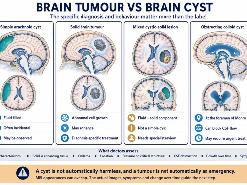
Choosing observation over immediate surgery is not the same as doing nothing. Structured follow-up with clinical review and imaging can be the safest course for a stable, asymptomatic lesion.

Do not find what you're looking for?

Our specialists are here to help. Get expert answers tailored to your condition—no waiting, no guesswork