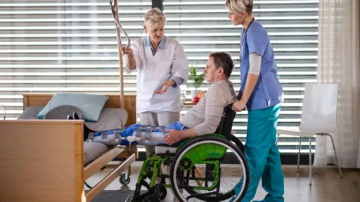
Drooling of Saliva

What is Drooling?

Drooling is the involuntary leakage of saliva from the mouth due to difficulty controlling facial muscles, lips, tongue, or swallowing mechanisms.

Common Causes

  • Bell's Palsy
  • Stroke
  • Parkinson's disease
  • Neurological disorders
  • Facial nerve injury 

Investigations

  • Neurological examination
  • Swallowing assessment
  • MRI Brain
  • Speech evaluation 

Treatment Options

  • Facial exercises
  • Speech therapy
  • Medications
  • Treatment of neurological condition 

Red Flags

  • Difficulty swallowing
  • Choking episodes
  • Stroke symptoms
  • Sudden onset 

FAQs

Can Bell's Palsy cause drooling?

Yes.

Is drooling a neurological symptom?

Often, yes.

Can therapy improve symptoms?

Yes.

When to Consult

Persistent drooling should be evaluated to identify underlying neurological or swallowing disorders.

Difficulty with Balance

What is Difficulty with Balance?

Difficulty with balance refers to an inability to maintain stability while standing, walking, turning, or performing routine activities. Patients may feel unsteady, dizzy, lightheaded, or as though they are about to fall. Balance depends on proper functioning of the brain, inner ear, spinal cord, nerves, muscles, and vision. Any disruption in these systems can lead to balance problems.

Balance issues can develop gradually or occur suddenly. In neurological conditions, they may indicate disorders affecting the cerebellum, brainstem, spinal cord, or peripheral nerves.

Common Causes

  • Concussion and traumatic brain injury
  • Stroke
  • Inner ear disorders
  • Parkinson's disease
  • Multiple sclerosis
  • Peripheral neuropathy
  • Cervical spine disorders
  • Brain tumors
  • Vestibular disorders
  • Medication side effects 

How is it Diagnosed?

A physician will evaluate:

  • Walking pattern (gait)
  • Coordination
  • Muscle strength
  • Sensation
  • Vision
  • Ear function
  • Neurological status 

Investigations

  • MRI Brain
  • CT Brain
  • Vestibular testing
  • Hearing assessment
  • Blood tests
  • Nerve conduction studies
  • Spine imaging 

Treatment Options

Treatment depends on the underlying cause and may include:

  • Balance rehabilitation
  • Vestibular therapy
  • Medications
  • Physiotherapy
  • Treatment of neurological conditions
  • Surgical intervention in selected cases 

When Should You Consult a Doctor?

Seek medical evaluation if balance problems:

  • Persist for several days
  • Cause falls
  • Interfere with daily activities
  • Occur suddenly 

Red Flags

  • Sudden inability to walk
  • Stroke symptoms
  • Severe dizziness
  • Limb weakness
  • Double vision
  • Loss of consciousness
  • Recent head injury 

FAQs

Can poor balance indicate a stroke?

Yes. Sudden balance difficulty can be an early sign of stroke.

Are balance problems related to ear disorders?

Yes. The inner ear plays a major role in maintaining balance.

Can physiotherapy help?

Yes. Specialized balance therapy often improves stability.

When to Consult:

Persistent balance problems should be evaluated by a neurology specialist to identify the cause and reduce the risk of falls and injuries.

Loss of Consciousness

What is Loss of Consciousness?

Loss of consciousness occurs when a person becomes unresponsive and unaware of their surroundings. It can last seconds, minutes, or longer.

Common Causes

  • Head injury
  • Concussion
  • Seizures
  • Stroke
  • Cardiac disorders
  • Metabolic abnormalities 

Investigations

  • CT Brain
  • MRI Brain
  • ECG
  • Blood tests
  • EEG 

Treatment

Emergency stabilization and treatment of the underlying cause.

Red Flags

All episodes require urgent medical assessment.

FAQs

Is brief unconsciousness serious?

It can be, especially after trauma.

Can concussion cause unconsciousness?

Yes.

Should I go to the hospital?

Yes.

When to Consult:

Loss of consciousness is a medical emergency and should never be ignored.

Stroke Symptoms

What is a Stroke?

A stroke (cerebrovascular accident) occurs when blood flow to a region of the brain is suddenly interrupted, either by arterial occlusion (ischemic stroke — approximately 85% of cases) or by rupture of a blood vessel (hemorrhagic stroke — approximately 15% of cases). Deprivation of oxygen and glucose leads to neuronal death; it is estimated that approximately 1.9 million neurons die per minute during an untreated ischemic stroke.

"Time is brain" — early reperfusion therapy with intravenous thrombolysis (tPA) within 4.5 hours and mechanical thrombectomy within 24 hours in selected patients significantly improves functional outcomes.

Powers WJ, et al. 2019 AHA/ASA Guidelines for the Early Management of Patients with Acute Ischemic Stroke. Stroke. 2019;50(12):e344–e418. doi:10.1161/STR.0000000000000211

The BE-FAST Acronym (Updated from FAST)

Recent evidence supports the expanded BE-FAST acronym to identify additional stroke symptoms often missed by FAST alone:

  • B — Balance: sudden difficulty with balance or coordination
  • E — Eyes: sudden vision change, double vision, or loss of vision
  • F — Face: facial drooping (ask patient to smile)
  • A — Arms: arm or leg weakness (ask patient to raise both arms)
  • S — Speech: slurred, confused, or no speech
  • T — Time: time to call emergency services immediately

Transient Ischemic Attack (TIA)

Stroke symptoms that resolve completely within 24 hours (and typically within 1 hour) may represent a Transient Ischemic Attack (TIA). TIA is NOT benign — it carries a high short-term risk of completed stroke (up to 10–15% within 90 days, with highest risk in the first 48 hours). All TIA patients require urgent evaluation and secondary prevention.

Johnston SC, et al. Transient ischemic attack: part 1. Diagnosis and evaluation. N Engl J Med. 2002;347(21):1714–1721. doi:10.1056/NEJMcp020190

Treatment

  • IV alteplase (tPA): eligible patients within 4.5 hours of symptom onset
  • Mechanical thrombectomy: eligible patients within 6–24 hours depending on imaging criteria
  • Blood pressure management per guidelines
  • Antiplatelet therapy (aspirin / dual antiplatelet in TIA)
  • Anticoagulation (in cardioembolic stroke/AF)
  • Stroke unit admission
  • Rehabilitation: physiotherapy, speech therapy, occupational therapy

🔴 RED FLAGS — Seek Emergency Care Immediately

• Any sudden onset of BE-FAST symptoms — call emergency services immediately

• Sudden severe headache (thunderclap) — may indicate subarachnoid hemorrhage

• Sudden loss of vision in one eye (amaurosis fugax)

• Stroke symptoms that resolve — still requires emergency evaluation (TIA risk)

Carotid Endarterectomy

Introduction:

Carotid endarterectomy (CEA) is a surgical procedure aimed at treating carotid artery stenosis, a condition characterized by the narrowing of the carotid arteries that supply blood to the brain. By removing the buildup of plaque in these arteries, CEA helps prevent further strokes and improve long-term outcomes. This article provides a comprehensive guide to CEA covering indications for surgery, preoperative preparation, intraoperative techniques, and postoperative care.


Image
Carotid


Primary goal of carotid endarterectomy

The primary goal of carotid endarterectomy is to prevent strokes by removing the plaque buildup in the carotid arteries. By restoring proper blood flow to the brain, CEA reduces the risk of future strokes and improves overall patient outcomes.


Indications for carotid endarterectomy

Carotid endarterectomy is indicated for patients with significant carotid artery stenosis (>50% narrowing according to NASCET) and a recent stroke or transient ischemic attack (TIA) related to that specific artery. Additionally, patients without symptoms but with severe stenosis (>70%) may also benefit from CEA based on individual assessment.


Patients Preparation for Carotid Endarterectomy

Preoperative preparation for carotid endarterectomy involves a thorough evaluation, which includes medical history review, physical examination, and imaging studies (such as carotid ultrasound, CT angiography, or magnetic resonance angiography). Medication management, particularly blood thinners, may be adjusted before surgery. It is essential to inform the surgical team about any allergies, medical conditions, or medications.


Role of Intraoperative Neuromonitoring during Carotid Endarterectomy

Intraoperative neuromonitoring is crucial during carotid endarterectomy to assess the integrity of the brain's blood supply and neurological function. Techniques such as electroencephalography (EEG), transracial Doppler ultrasound, or somatosensory evoked potentials (SSEPs) are used to monitor the brain's electrical activity and blood flow during the surgery. This monitoring helps ensure the safety of critical neurological structures.


Procedure Performed, and What Role does the Microscope Play

Carotid endarterectomy is typically performed under general anesthesia. An incision is made along the front of the neck, and the carotid artery is exposed. The plaque is carefully removed from the artery, and the vessel is repaired with stitches or a patch. A surgical microscope is commonly used during the procedure to provide a magnified view, improving precision and minimizing the risk of damage to surrounding structures.


Carotid Endarterectomy differ from Carotid Stenting

Carotid endarterectomy and carotid stenting are two different approaches to treat carotid artery stenosis. CEA involves the surgical removal of plaque, while carotid stenting involves the placement of a stent to keep the artery open. The choice between the two procedures depends on various factors, including the patient's overall health, anatomy, and surgeon's expertise.


Postoperative Care and Long-term Outcomes

After carotid endarterectomy, patients are closely monitored in the postoperative intensive care unit (ICU) for a short period. Vital signs, neurological status, and incision site are regularly assessed. Blood thinners or antiplatelet medications may be prescribed to prevent clotting. Long-term outcomes of carotid endarterectomy are generally favorable, with a reduced risk of recurrent stroke and improved overall prognosis.


Conclusion:

Carotid endarterectomy is a vital surgical procedure for the treatment of carotid artery stenosis, aimed at preventing strokes and improving long-term outcomes. Indications for CEA include significant stenosis, recent stroke or TIA, and individual assessments. Preoperative preparation, intraoperative neuromonitoring, and the use of a microscope contribute to the safety and precision of the procedure. Distinct from carotid stenting, CEA involves the surgical removal of plaque. Postoperative care involves monitoring in the ICU, medication management, and follow-up evaluations. With careful patient selection and appropriate surgical intervention, carotid endarterectomy has shown to be effective in reducing the risk of recurrent stroke and improving long-term outcomes for patients.


Subscribe to stroke